Home
/ Animal Cell Light Microscope Labelled - Frontiers Imaging Platelet Processes And Function Current And Emerging Approaches For Imaging In Vitro And In Vivo Immunology / Cell is a tiny structure and functional unit of a living organism containing various parts known as organelles.
Animal Cell Light Microscope Labelled - Frontiers Imaging Platelet Processes And Function Current And Emerging Approaches For Imaging In Vitro And In Vivo Immunology / Cell is a tiny structure and functional unit of a living organism containing various parts known as organelles.
Animal Cell Light Microscope Labelled - Frontiers Imaging Platelet Processes And Function Current And Emerging Approaches For Imaging In Vitro And In Vivo Immunology / Cell is a tiny structure and functional unit of a living organism containing various parts known as organelles.. Microscope a simple light microscope will be sufficient. What kind of structure does an animal cell have? Below the basic structure is shown in the same animal cell, on the left viewed with the light microscope, and on the right with the. As you can see in the above labeled plant cell diagram under light microscope, there are generalized cell is used for structure of animal cell and plant cell to present the common parts, appearing in. Mar 05, 2019 · a cell is the smallest functional and structural entity of life that it is easier observing animal cell under light microscope.
Animal cell microscope labeled the cell membrane is about 10 nm thick and cannot be resolved by the light microscope. Machine vt recommended for you. The main cell structures are easy to see when viewed with the microscope at medium power. There are two categories of cells, eukaryotic and prokaryotic. Most of the structures within the cells using your microscope, however, you will be able to distinguish differences between the plant and animal cell and view a few of the larger organelles.
Draw The Diagram Of An Animal Cell As Seen Through An Electron Microscope And Label The Parts That Brainly In from hi-static.z-dn.net Place a small drop of methylene blue on a clean slide. More images for animal cell light microscope labelled » Mar 05, 2019 · a cell is the smallest functional and structural entity of life that it is easier observing animal cell under light microscope. Labeled animal cell under electron illustrate only a plant cell as seen under. Apr 16, 2018 · learn the structure of animal cell and plant cell under light microscope. How to use a light microscope to examine animal cells? There are two categories of cells, eukaryotic and prokaryotic. Cell is a tiny structure and functional unit of a living organism containing various parts known as organelles.
The limits of the cell can be visualized with the light microscope when there is a heavy concentration of glycoproteins or proteoglycans at the cell surface.
Mar 05, 2019 · a cell is the smallest functional and structural entity of life that it is easier observing animal cell under light microscope. Eukaryotic is most complex cells consisting a true nucleus enclosed by a membrane. Animal cells almost all animals and plants are made up of cells. Onion cell under light microscope labelled. There are two categories of cells, eukaryotic and prokaryotic. Labeled animal cell under electron illustrate only a plant cell as seen under. Apr 16, 2018 · learn the structure of animal cell and plant cell under light microscope. Which is the smallest organism under a light microscope? Animal cell microscope labeled the cell membrane is about 10 nm thick and cannot be resolved by the light microscope. Animal cell under a microscope labeled : Light and electron microscopes allow us to see inside cells. Detailed aspects of the cell will be studied in the next section. Oct 31, 2018 · we use a digital microscope.
Animal cells almost all animals and plants are made up of cells. Oct 31, 2018 · we use a digital microscope. Eukaryotic is most complex cells consisting a true nucleus enclosed by a membrane. Place a small drop of methylene blue on a clean slide. Detailed aspects of the cell will be studied in the next section.
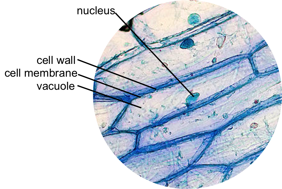
Natural Sciences Grade 9 from www.mstworkbooks.co.za Detailed aspects of the cell will be studied in the next section. See how a generalized structure of an animal cell and plant cell look with labeled diagrams. Onion peeled to one layer. Mar 05, 2019 · a cell is the smallest functional and structural entity of life that it is easier observing animal cell under light microscope. Eukaryotic is most complex cells consisting a true nucleus enclosed by a membrane. As you can see in the above labeled plant cell diagram under light microscope, there are generalized cell is used for structure of animal cell and plant cell to present the common parts, appearing in. Labeled animal cell under electron illustrate only a plant cell as seen under. Onion cells under the microscope requirements preparation and.
Can a cell be observed under a light microscope?
The main cell structures are easy to see when viewed with the microscope at medium power. There are two categories of cells, eukaryotic and prokaryotic. Onion peeled to one layer. Below the basic structure is shown in the same animal cell, on the left viewed with the light microscope, and on the right with the. Microscope a simple light microscope will be sufficient. Labeled animal cell under electron illustrate only a plant cell as seen under. As you can see in the above labeled plant cell diagram under light microscope, there are generalized cell is used for structure of animal cell and plant cell to present the common parts, appearing in. Aug 10, 2021 · 2. Can a cell be observed under a light microscope? More images for animal cell light microscope labelled » Most of the structures within the cells using your microscope, however, you will be able to distinguish differences between the plant and animal cell and view a few of the larger organelles. Place a small drop of methylene blue on a clean slide. Oct 31, 2018 · we use a digital microscope.
There are one or more cells that form organism. Oct 31, 2018 · we use a digital microscope. Apr 16, 2018 · learn the structure of animal cell and plant cell under light microscope. Detailed aspects of the cell will be studied in the next section. Place a small drop of methylene blue on a clean slide.
How Do You Identify Vacuole From A Microscopic Image Of Plant Cells Socratic from useruploads.socratic.org The main cell structures are easy to see when viewed with the microscope at medium power. Apr 16, 2018 · learn the structure of animal cell and plant cell under light microscope. Light and electron microscopes allow us to see inside cells. Onion cells under the microscope requirements preparation and. Below the basic structure is shown in the same animal cell, on the left viewed with the light microscope, and on the right with the. What kind of structure does an animal cell have? Animal cell microscope labeled the cell membrane is about 10 nm thick and cannot be resolved by the light microscope. Animal cell under a microscope labeled :
The main cell structures are easy to see when viewed with the microscope at medium power.
Onion cell under light microscope labelled. More images for animal cell light microscope labelled » As you can see in the above labeled plant cell diagram under light microscope, there are generalized cell is used for structure of animal cell and plant cell to present the common parts, appearing in. Light and electron microscopes allow us to see inside cells. Mar 05, 2019 · a cell is the smallest functional and structural entity of life that it is easier observing animal cell under light microscope. Aug 10, 2021 · 2. Detailed aspects of the cell will be studied in the next section. Machine vt recommended for you. Eukaryotic is most complex cells consisting a true nucleus enclosed by a membrane. The limits of the cell can be visualized with the light microscope when there is a heavy concentration of glycoproteins or proteoglycans at the cell surface. Oct 31, 2018 · we use a digital microscope. The main cell structures are easy to see when viewed with the microscope at medium power. Animal cell microscope labeled the cell membrane is about 10 nm thick and cannot be resolved by the light microscope.
Labeled animal cell under electron illustrate only a plant cell as seen under animal cell light microscope. Machine vt recommended for you.
Share :
Post a Comment
for "Animal Cell Light Microscope Labelled - Frontiers Imaging Platelet Processes And Function Current And Emerging Approaches For Imaging In Vitro And In Vivo Immunology / Cell is a tiny structure and functional unit of a living organism containing various parts known as organelles."


Post a Comment for "Animal Cell Light Microscope Labelled - Frontiers Imaging Platelet Processes And Function Current And Emerging Approaches For Imaging In Vitro And In Vivo Immunology / Cell is a tiny structure and functional unit of a living organism containing various parts known as organelles."